As a long-time student of invertebrate zoology I have for most of my life appreciated the immense variety and ingenuity of animal body plans. Most of the animals that we are familiar with (think of any pets you’ve ever had) have what’s called bilateral symmetry: they have a head end and a tail end, a left and a right, and a top and a bottom. In scientific terms that translates to the anterior-posterior, left-right, and dorsal-ventral axes. Also, most bilateral animals are elongated on the anterior-posterior axis and have some sort of cephalization going on in the anterior end of the body. In other words they have a head, or at least a concentration of neural tissue and sensory structures in the part of the body that encounters the environment first.
Even your basic worm meets all these criteria. Here’s a video clip of Alitta sp., an intertidal polychaete worm. The polychaetes are somewhat distant relatives of earthworms, and both groups have the distinctly segmented body plan of their phylum, the Annelida. Alitta’s body symmetry is clearly bilateral and you can see that it has an anterior end, which in this case is defined by both the direction of locomotion and the presence of a head:

As “normal” as bilateral symmetry may seem—and it seems normal to us only because it is the way of our own bodies—there are many animals whose bodies are organized in completely different ways. The cnidarians, for example, are the largest group of animals with radial symmetry. Instead of being elongated along an anterior-posterior axis, these animals’ bodies are either columnar or umbrella-shaped. In either case, when you look down on them you see a circular shape:
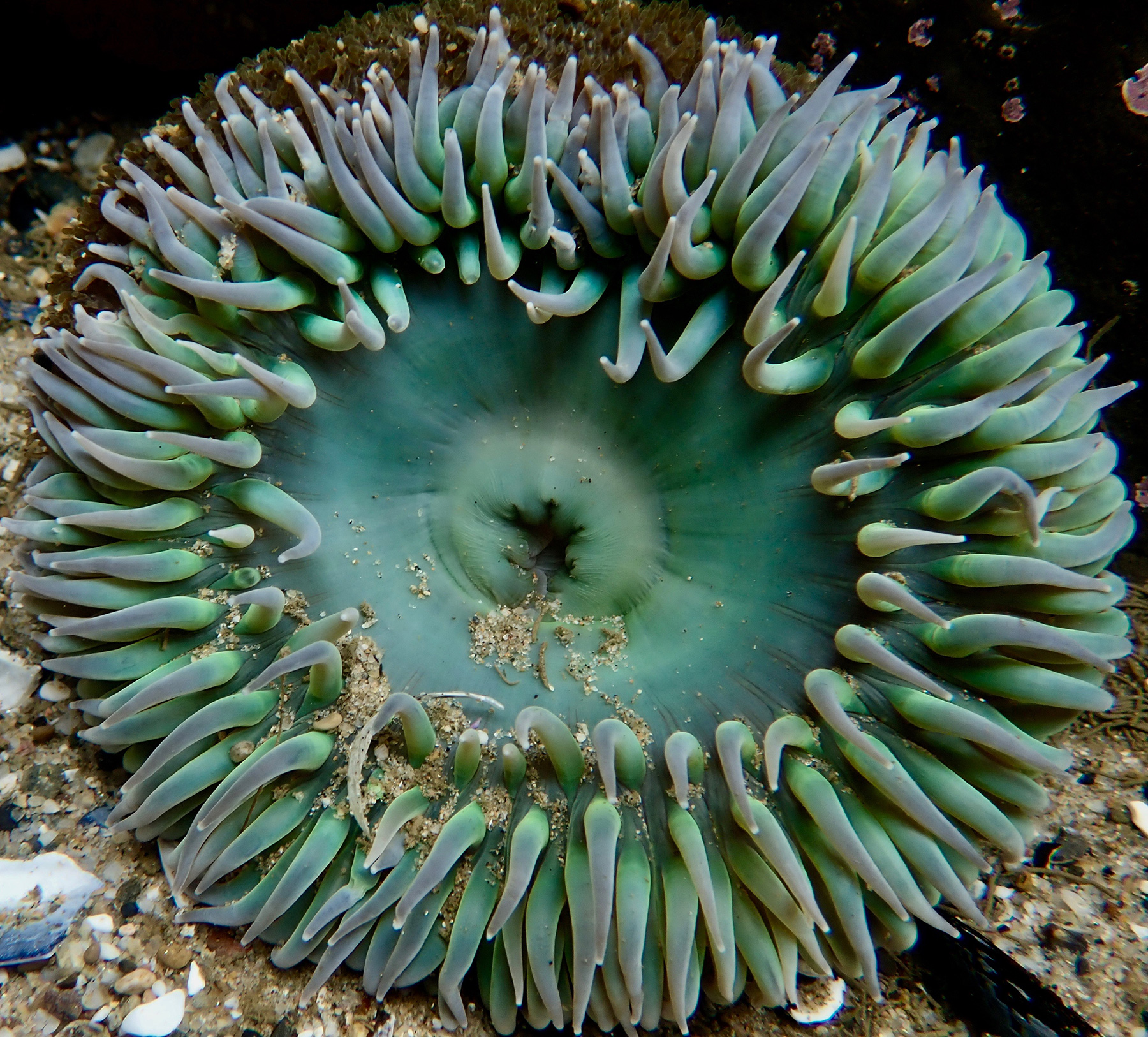
An animal with this sort of body plan obviously has no head–no eyes, nose, or concentration of either neural or sensory structures. Being a sea anemone, it lives attached to the sea floor and doesn’t walk around much, so we can’t take hints about front and back from the direction of its movement like we could with the polychaete worm. Rather than have most of the neural apparatus concentrated in any particular region, a sea anemone’s nervous system is diffusely scattered over the entire body. This animal has the advantage of meeting its environment from all sides and across the entirety of its external surface. It can respond to the presence of food or the approach of a predator from any direction. It can’t be sneaked up on, because it has no front or back.
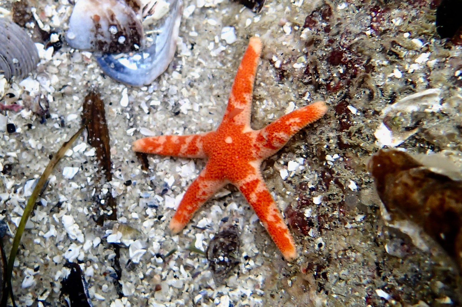
The echinoderms are another group of animals with radial symmetry. The phylum Echinodermata comprises sea stars, sea urchins, sand dollars, brittle stars, sea cucumbers, and crinoids. Echinoderms’ bodies are indeed radial in nature, but in a different way from the radial symmetry of cnidarians’ bodies. The general echinoderm body plan is organized around the number five, so we say that they have pentaradial or pentamerous symmetry.
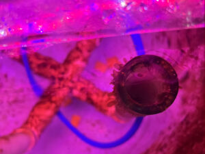
To make sense of this pentaradial symmetry, let’s take a look at the defining characteristic of echinoderms, the water vascular system. This internal structure consists of a series of fluid-filled tubes and canals, connected to the exterior of the body by a calcareous plate called the madreporite. In many species of sea stars, such as the ochre star in the photo below, the madreporite is visible as a flattened granular structure located off-center on the aboral (top) surface.
Seawater flows through the madreporite to maintain hydraulic pressure within the water vascular system. In the center of the animal, surrounding the esophagus, is a circular tube called the ring canal. Emanating from the ring canal are five radial canals, from which protrude the suckered tube feet that are used for feeding and/or locomotion. As you might imagine, the five-way symmetry of echinoderms has strong implications both for other aspects of the animal’s anatomy and the way that it interacts with its environment.
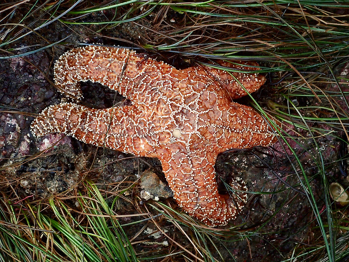
Echinoderms are structurally more complex than cnidarians, with distinct internal organs. The central disc contains most of the organs, but there are extensions of both the gut and the gonads in each of the five arms. Like the cnidarians, the echinoderms don’t have a centralized nervous system, but they do have very simple eyes that can detect light and dark. And guess where, in a sea star, the eyes are located? Hint: Think about how the animal encounters its environment. Yes, the eyes are in the tips of the arms, along with chemosensory receptors. Makes sense, doesn’t it?
Compared to the sedentary or sessile cnidarians, echinoderms are very active, and most of them can crawl around on the benthos. As expected, their pentaradial symmetry affects how they move. Having no front or back, sea stars and sea urchins can walk in any direction they want—towards food or away from a predator, for example. They can also change the direction of locomotion easily, without needing to turn around the way we would.
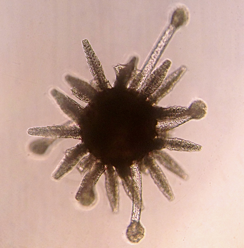
Pentaradial symmetry doesn’t occur in any animal group except the echinoderms, and even they begin life as bilateral larvae. This is the pluteus larva of the purple sea urchin, Strongylocentrotus purpuratus, age 31 days post-fertilization:
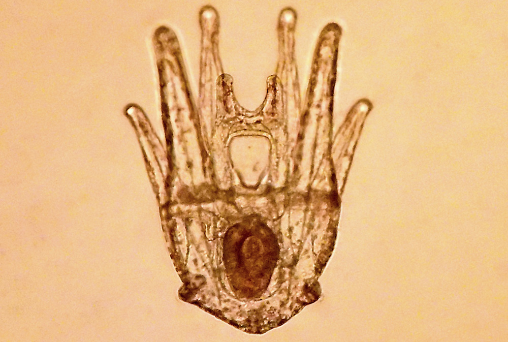
There isn’t a more perfect example of bilateral symmetry out there. Although, even at this stage there are developments within the body that are beginning to interrupt the bilateral-ness of the animal. This is a picture of the animal lying on its front, so you are looking down on its back. At this age the pluteus larva has four pairs of arms, each of which is supported by an internal skeletal rod. The clear structure in the center, which looks like an upside-down milk jug and sits just above the horizontal midline, is the mouth. An esophagus leads to the stomach, which is that oblong darker structure in the center of the body. A continuous ciliated band runs up and down all eight of the arms, and is just barely visible as a slight pale halo around the edges of the arms. The beating of the ciliated band propels the pluteus through the water and also brings food to the mouth.
See how, to the upper left of the stomach, at about 11 if it was the face of a clock, there is a darkish squiggle running mostly horizontally between the stomach and the skeletal rod of that arm, that you don’t see on the right side? That squiggle indicates where the juvenile rudiment, which contains the first five tube feet of the water vascular system, will develop. This rudiment grows to the point that it occupies most of the internal space of the pluteus. As the rudiment grows, the body of the pluteus gets heavy. Eventually the larva reaches a stage called competence, which means that it is anatomically and physiologically ready to fall out of the plankton, metamorphose into the juvenile body, and begin life on the benthos. When the larva settles out of the plankton it lands on its left side, where the tube feet erupt through the body wall during metamorphosis. The end result is a little urchin walking around on tube feet that it didn’t have the day before. (Well, I guess technically it had them, but there weren’t useful yet.) And the body symmetry will have changed from the bilateral larval form to the pentaradial juvenile.
When looking at a live sea urchin it can be difficult making sense of all the stuff that’s going on. A sea urchin is a very active animal, with spines and tube feet waving all over the place. It looks like total chaos, but examination of a naked sea urchin test (the endoskeleton made up of interlocking calcareous ossicles) lends a lot of insight into the body plan of this animal.
Here’s a cleaned intact urchin test:
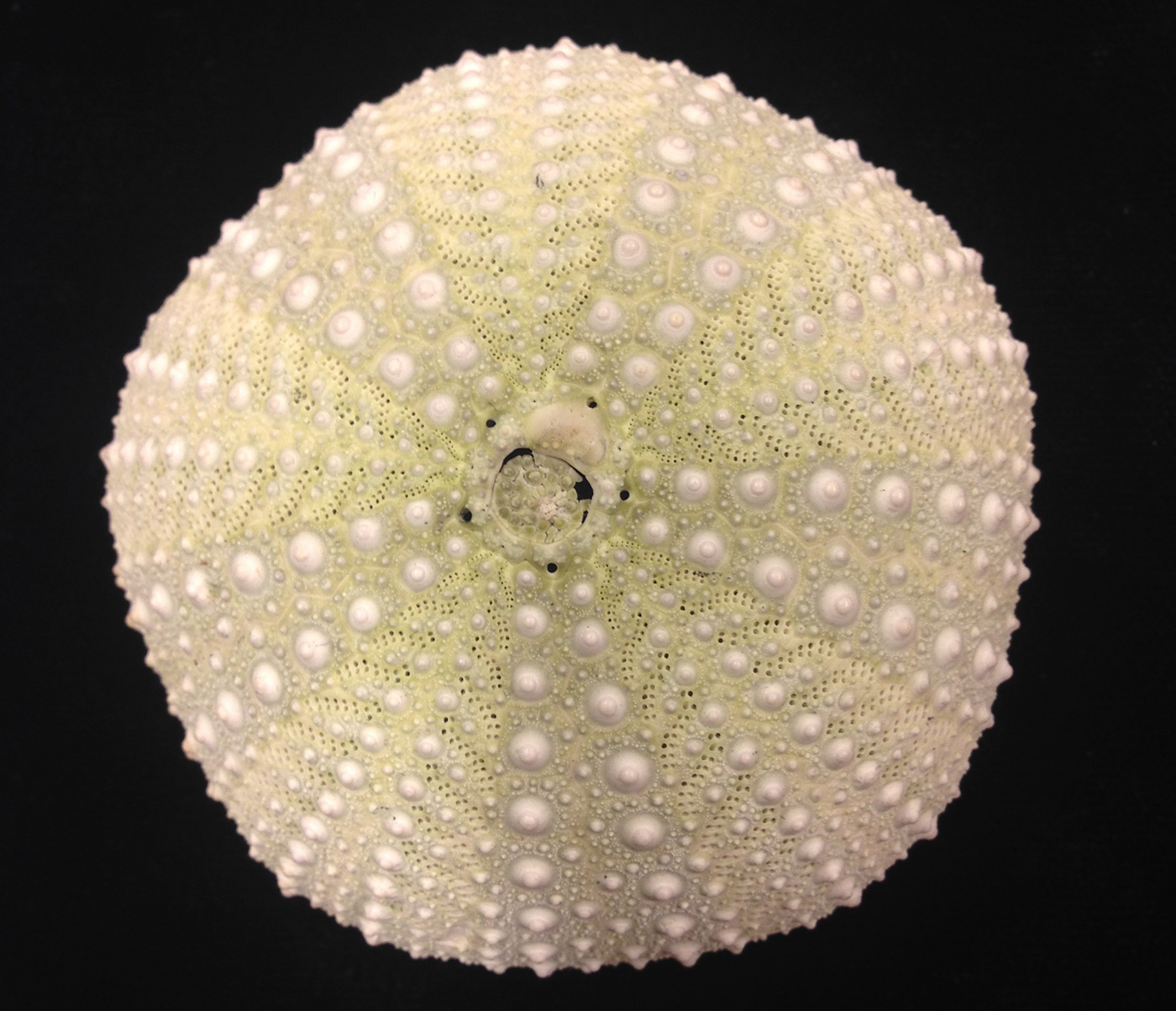
Now the pentaradial symmetry of this body plan becomes apparent. You can see that there are five regions of doubled rows of plates that have little holes in them. The holes are where the tube feet protrude to the outside, and the plates that bear them represent the animal’s ambulacrum, or ambulacral region. The radial canals of the water vascular system run up along the inside surface of the test in the five ambulacra. The ambulacral regions are separated from each other by five intermabulacral regions, which do not have holes for tube feet because there are no tube feet here. The bumps on the test are called tubercles, and are where the spines attach. The tubercles fit into the base of the spines like a ball-and-socket joint, similar to our shoulder, which allows the spines to rotate 360 degrees. You can see this for yourself the next time you have a live urchin available: touch one of the spines and observe how the animal reacts.
There is interesting stuff going on at the apex of the urchin, too. The five larger holes, one at the point of each interambulacral area, are the gonopores. When I inject urchins with magnesium chloride to induce spawning, the gametes are released from these holes. The arrangement of the gonopores in the interambulacral regions makes sense, once you remember that on the inside of the test the ambulacral areas are where the water vascular system structures (including tube feet) are located. The only space available for the gonads is in the interambulacral areas. I know, it’s confusing. And people think invertebrates are simple!
There’s a natural human tendency to regard creatures like us as somehow better than those different from us. We certainly do display more empathy for mammals than we do for, say, insects or worms. I teach my students that complex is not always better (think of the pervasive damage done to a person who has suffered a major brain or spinal cord injury) and that there are multiple types of complexity—regarding morphology, behavior, reproduction, and life cycle—in the animal kingdom. The best way to understand an animal is to put yourself in its “shoes” and try to imagine what its life is like, with its anatomy, physiology, and lifestyle. For example, pretend that you are a sea star. You have no head, crawl around on your belly, and have eyes at the ends of your limbs. How would you experience the world if you had this body? In what ways would it be different from your life as a human being? It takes some mental gymnastics and can be difficult to shed our human-centric biases, but we must be able to put them aside at least temporarily if we truly want to make sense of what’s going on in the world around us.